Our services
Plateforms, Animal models and Technology
  • Renal function
  • Blood flow assessment and limb function
  • Atherosclerosis
  • Cre-Lox system
  • Live Cell Imaging
Renal function

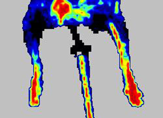
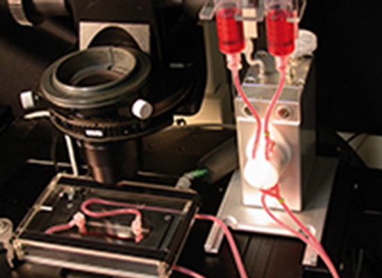
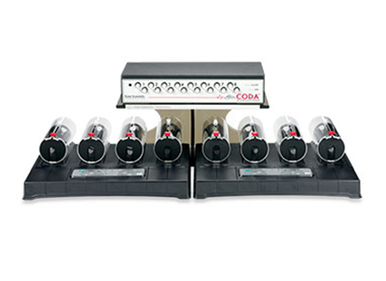
In our different animal models, we have the capacity to evaluate renal function by measuring arterial pressure with the non-invasive tail-cuff system, glomerular filtration rate (GFR) and albuminuria (ELISA).

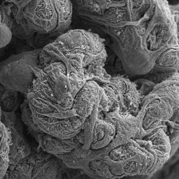
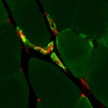
The structure and the integrity of the podocytes can be analyzed with transmission and scanning electronic microscopy in addition to histopathological analyses of the tubules and glomeruli.

Blood flow assessment and limb function

Using the lower limb ischemia model, blood flow measurement can be performed with the laser Doppler PIMIII (Perimed).In addition, our laboratory can perform limb motricity using voluntary running wheels.

Following several weeks of monitoring, histological and immunofluorescence analyses of the muscle are performed for capillary density.

Atherosclerosis

Using pro-atherogenic mouse models (apoE and LDLr deficient mice), our laboratory is investigating the interactions of circulating cells (monocytes) with the endothelium that occur during the development and progression of the atherosclerotic plaque in diabetes. Moreover, our laboratory can perform bone marrow transplant to evaluate monocyte contribution to atherosclerotic plaque formation.

Cre-Lox System

We have a variety of conditional Cre-Lox models to target specific genes that are involved in podocyte, endothelial and smooth muscle cells.

We have recently generated a transgenic mouse using the FLEX-Cre-Lox technology.

Live Cell Imaging

With the financial support of the Canadian Foundation for Innovation (CFI), we have the capacity to visualize in real-time the structure, the morphology and cell-to-cell interactions caused by diabetes.

Publications
Recent published articles
  • By Benoît Denhez
  • May 1st, 2019

Reduction of DUSP4 in podocytes.

We showed for the first time that diabetes reduces DUSP4 expression in renal podocytes, which causes glomerular fibrosis and diabetic nephropathy.

SHP-1 and smooth muscle cell function in diabetes.

Diabetes maintained SHP-1 activity preventing hypoxia-induced PDGF actions in smooth muscle cells. Specific deletion of SHP-1 in SMC partially restored blood flow reperfusion in the diabetic ischemic limb.

Transcriptomic monocyte profile following high-intensity interval training in diabetes.

The downregulation of transcripts in monocytes globally suggests a favorable cardiovascular effect of the high-intensity interval training in older women with type 2 diabetes. In the context of precision medicine and personalized exercise prescription, shedding light on the fundamental mechanisms underlying HIIT effects on the gene profile of immune cells is essential to develop efficient nonpharmacological strategies to prevent cardiovascular disease in high-risk population.

Funding sources and partners